Researchers from Vanderbilt University and Vanderbilt University Medical Center have received a $1.4 million grant from the National Eye Institute at the National Institute of Health to identify surgical techniques that improve vision after macular hole repair.
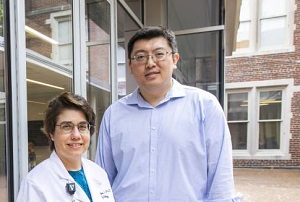
Yuankai “Kenny” Tao, assistant professor of biomedical engineering, is the principal investigator and leads a team of engineers and clinicians that includes Karen Joos and Shriji Patel, both ophthalmologists at Vanderbilt Eye Institute. All study investigators are affiliates of the Vanderbilt Institute for Surgery and Engineering as well.

Macular holes are tears in the retina that can blur or distort vision. The disease is age-related and usually occurs in people over 60. When such holes occur in the central vision, patients can lose the ability to see fine details, read or drive. Left untreated, macular holes can lead to retinal detachments and loss of vision. Severe macular holes are routinely repaired surgically, however, how much vision is restored after surgery varies highly from patient to patient.
Researchers want to translate technologies that enable imaging of macular holes during surgical repair. They believe that differences in vision restoration after surgery is directly related to the integrity of the retina during surgery. By imaging retinal layers, the team hopes to better understand how different surgical techniques impact retinal healthy and correlate changes in retinal structure during surgery with visual outcomes after surgery.
The outcomes of this study will allow for more precise surgical planning and better prediction of visual outcomes after macular hole repair, Tao said. Understanding how surgical techniques affect retinal integrity can also help development novel surgical techniques, he said.