As reported by the National Institute of Biomedical Imaging and Bioengineering (NIBIB)
For patients with movement disorders such as Parkinson’s disease, simple activities such as drinking a cup of coffee or walking to the dinner table present a challenge. Their limbs jerk or move without control. Medication can ease these symptoms, but over time the body stops responding to drug therapy.
Many of these cases are helped by deep brain stimulation (DBS), a surgical procedure in which electrodes are placed in the brain at key sites responsible for movement. Wires attached to the implants are secured to an electrical stimulation device similar to a heart pacemaker that is placed under the skin near the collarbone. Electrical pulses, which travel from the stimulator along the lead wire to the implant, block abnormal nerve signals that cause tremors and other unwanted movements.
When approved by the U.S. Food and Drug Administration 12 years ago, DBS surgery could take up to 12 hours. After drilling holes in the skull, a surgeon would estimate the best trajectory for threading the electrode through the brain’s anatomy to the target site. However, because each patient’s brain is slightly different and structures may be a few millimeters to the left or right, the surgeon sometimes needed six to ten attempts to hit the optimal target. Patients were awake during the surgery so that they could provide feedback to the surgeon. For older patients, the lengthy procedure could be exhausting. Today, advances in technique and equipment have shortened surgery times to just a few hours.
Engineers and a neurosurgeon at Vanderbilt University are testing a knowledge repository and interactive software that have improved the accuracy of implant placement and, thereby, cut surgery time to as little as two hours.
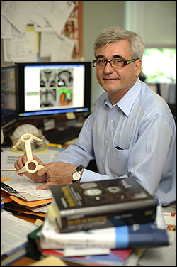
 Benoit Dawant |
The database known as CranialVault and its Cranial Vault Explorer (CRAVE) software tools result from a nearly decade-long collaboration between Vanderbilt neurosurgeon Peter Konrad and Benoit Dawant, professor of electrical engineering and biomedical engineering, and Pierre-Francois D’Haese, a research assistant professor in electrical engineering and computer science.
Though a number of tools developed over the last decade have helped surgeons in various aspects of the DBS process, CranialVault and CRAVE are the first tools to span the entire spectrum of the procedure, from preoperative planning to surgical implantation and device programming.
By combining the database and interface tools, the researchers say they’ve created a system that could become a national repository for data from many sites around the country, eventually allowing them to compare best practices across sites.
“If we do our job right, we will be able to capture the experiences of many surgeons and enable others to benefit from that experience,” says Dawant, principal investigator on the project.
Building the Vault
The heart of the system, the CranialVault database, stores information on patients who have undergone the DBS procedure. The data include computed tomography (CT) scans and magnetic resonance imaging (MRI) studies, preoperative surgical plans, position of the final implants, as well as data points from microelectrode recordings used during surgery to distinguish various neuronal firing patterns. The database also holds data showing a patient’s response to stimuli during surgery.
Currently, the CranialVault – which, along with the CRAVE tools, is secure and HIPAA compliant – contains records from more than 450 patients. A further strength of the CranialVault is its ability to create brain maps based on information from all patients in the database.
 Pierre-Francois D’Haese |
Researchers use statistical data from all of the patients in the CranialVault database to build a sort of “mega-brain” or map of the average brain. Applying mathematical equations or algorithms first published by D’Haese in 2003, researchers project data from one brain onto the mega-brain. The result is a statistical map showing the anatomy of each brain relative to those of other patients. These maps are then used to plan surgeries, locate optimal implant sites, and program the stimulator.
Mining the Vault
Other algorithms are dedicated to the robust user interface called CRAVE. This suite of tools allows the DBS team to garner specific information about the patient during each stage of the DBS process. In the surgery planning phase, the new patient’s images are added to the CranialVault and then morphed to the existing atlases. The system generates a preoperative plan based on targets chosen by the surgeon through the CRAVE planning module on a secure Web site. The program predicts the location of key brain structures in the trajectory and provides maps showing optimal implant locations for best outcomes and minimal side effects.
“We can project data from 100 successful surgeries onto a new patient’s brain and know that it is highly likely that if you implant at a particular location you’ll get a successful outcome,” says Dawant. So far the atlas has predicted site selections to within 1 mm for placement in the subthalmic nucleus, a common target site.
The DBS team uses CRAVE as a visual guide throughout surgery. The team can view the trajectories from the presurgical plan, as well as maps of the brain’s anatomy and statistical maps showing the best locations for electrode placement. CRAVE also provides three-dimensional (3-D) images of nerve activity to help surgeons find the best location to begin stimulation testing. Without the 3-D images, surgeons must create their own mental images based on a series of data points. Because CRAVE permits recording of electrical stimulation data, the team can use the 3-D images for final placement of the implants.
“Before we had the program, each patient’s surgery was like starting from ground zero,” says Konrad. To determine the optimal location for the DBS implant, the surgeon had to find where the patient’s recordings and response to electrical stimuli were best. “Each additional pass to gather more information raises the risk and impacts the patient’s performance as time passes in the operating room. With the software tied to the database, we have reduced the number of passes we make in the operating room because we tend to land right on the target more quickly.” DBS surgery risks include bleeding in the brain, which could lead to stroke or death, spinal fluid leakage, and infection. Recently, Konrad, who describes the program as “transforming,” placed electrodes in the brain of a 75-year-old professor in about 30 minutes and completed the entire surgery in less than 2 hours.
The program’s precision is especially helpful when surgeons encounter brain asymmetries, structures that do not match up exactly on each side of the brain. Konrad tells of a case in which the computer atlas suggested a target site 5 mm away from where he thought the target should be. He chose not to follow the program’s recommendation because it appeared too great a deviation from the expected site location. Once inside the brain, he found the atlas had correctly predicted the optimal site and he had to adjust the electrode placement to match the atlas’ suggested site.
Transforming Programming With CRAVE
In the programming stage of DBS, neurologists determine the correct electric voltage and the frequency of the pulses produced by the stimulator device to relieve patient symptoms. To do this, neurologists test each of four contacts on an implant to find the one that best eliminates symptoms, relying on tabular data from the patient’s prior electro-stimulation tests. Working through all four contacts can take several hours as different voltages and frequencies are tried; unfortunately, insurance pays for only two hours of testing.
Using CRAVE, programming becomes a visual process, with the location of the implants and results from electrical stimulation performed during surgery superimposed on the patient’s MRI. The system also predicts which contact is the best choice to activate based on CranialVault data. “If you can see where the lead is positioned and can draw on experience from previous patients, programming becomes much easier,” says Konrad. “The visual display will dramatically change the way DBS programming is done.”
With device manufacturers considering development of implants with 8, 16, or more contacts to improve the shape of the electrical field at the target site, Konrad notes that visualization software will become a requirement for DBS programming.
CRAVEing the Future
As the researchers continue to refine the CranialVault and CRAVE interface tools, Dawant says they will focus on integrating the system from planning to programming. “We want to create a pipeline to move the data through the entire clinical process,” says Dawant. Although the planning and intra-operative modules are mature, the programming phase has been used only sporadically. Programs to generate postoperative data sets for each surgical patient are under development, as are assessment tools to determine the programming module’s value for neurologists.
The team will also create new software to address changes in brain position during surgery. Unlike other brain surgeries in which large incisions expose much of the brain, access during DBS surgery only requires small burr holes drilled in the skull. Any time the brain is exposed – even through small holes – the brain shrinks, and this change can alter the position of brain structures. Measuring just how much the brain shrinks during DBS surgery is a challenge says Dawant. “[To devise a tracking model] we need to know how much the cortical surface moves, but with the small burr holes it’s difficult to determine,” he says.
The group also will continue collaborating with other large DBS centers to extend and develop new user interface tools, not only for movement disorders but also for conditions such as epilepsy and depression. Says Konrad, “We’re thinking about DBS in the next 10 years and what tools people will need to advance the field.”
This research is supported in part by the National Institute of Biomedical Imaging and Bioengineering and the National Institute of Neurological Disorders and Stroke (NINDS).