A new device that can image diseases of the retina more quickly will soon be tested during ophthalmic surgeries with Vanderbilt Eye Institute collaborators.
The prototype was designed by a Vanderbilt engineering undergraduate, who is first author on a paper about the work she will present today at the largest photonics conference in the world.
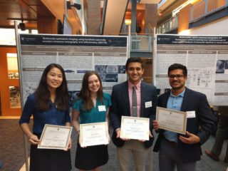
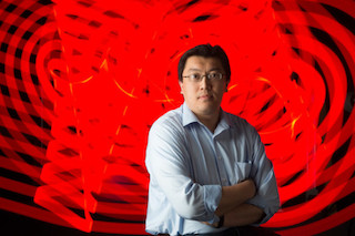
Kelsey Leeburg, a junior planning to double major in biomedical and electrical engineering, is one of four undergraduates presenting their work at SPIE Photonics West. All four worked last summer in the Diagnostic Imaging and Image-Guided Interventions Laboratory (DIIGI) under the direction of Yuankai “Kenny” Tao, assistant professor of biomedical engineering.
Leeburg; Oscar Benavides, a biomedical engineering major; Xiaoyue (Judy) Li; and Benjamin Terrones, a biomedical engineering senior, all had their conference abstracts weighed against those from graduate students, post-doctoral scholars and principal investigators.

“SPIE does not distinguish abstract submissions/invitation for presentations based on education level,” Tao said. “The subconferences they’re presenting in are particularly rigorous in reviewing abstracts because of the number of submissions they receive.”
The School of Engineering sent dozens of students, postdocs, staff researchers and professors to San Francisco for the conference, which wraps up later this week. Tao’s lab, which is affiliated with the Vanderbilt Institute for Surgery and Engineering, has six presentations, including the four by undergraduates; the Vanderbilt Biophotonics Center, led by Orrin H. Ingram Professor of Biomedical Engineering Anita Mahadevan-Jansen,
has 16 in all. The Vanderbilt team also includes Cornelius Vanderbilt Professor of Engineering Sharon Weiss, a professor of electrical engineering and computer science, and Dr. Karen Joos, Joseph and Barbara Ellis Professor of Ophthalmology.
Leeburg had the fifth highest-ranked abstract in the Optical Coherence Tomography (OCT) sub-conference. She designed and assembled a handheld spectrally encoded coherence tomography and reflectometry (SECTR).
“I am still amazed at how much I learned this summer, given that I knew very little about optics at the beginning,” she said. “I can now build a spectrometer and OCT system as well as explain how they work.
“I was excited about being able to work with my hands so much,” Leeburg said.
Li presented Saturday, Jan. 27, on using OCT and fluorescence confocal SLO (cSLO) to look at vascular changes in zebrafish retina.
Benavides, who built a combined cSLO-OCT imaging system to monitor murine injections for ocular research, and Terrones, who used multimodal OCT and cSLO to guide eye injections for ophthalmic research in mice, presented Sunday.
“For me it was interesting partly because it was completely new,” said Terrones, whose project received special recognition in September from the Vanderbilt Undergraduate Summer Research Program.
“I did not have much exposure to optics or imaging prior to joining the lab, and the challenge of learning all this new information was exciting. Additionally, the process of designing a novel system and having some creative freedom was also appealing,” he said.
The 10-week summer research posts were funded through VISE, the School of Engineering, and Tao’s lab. The student work, like the rapidly growing field itself, is multidisciplinary and collaborative. Each project involved other researchers, including graduate students and other undergrads, and the work encompassed biomedical engineering, electrical engineering, imaging and computational science.

“Our work is very focused in the eye and spans multiple species to allow us to tackle different parts of the problem,” Tao said. “The animal work is used to identify disease mechanisms and novel therapeutics, whereas the human imaging work is for early diagnostics and translating novel therapies.”
Several applications motivated development of a handheld device with technology that allows faster imaging and track motion to cancel it out in post-processing.
“We’re starting to get into imaging premature infants with retinopathy of prematurity. They can’t fixate and obviously can’t upright, so we really need a handheld device for this,” Tao said. “Similarly, this is useful for imaging the elderly and bedridden. There are applications in field-deployable devices for combat situations as well.”
The conference attracts more than 20,000 attendees in photonics, laser, and biomedical optics research and industries. The technical conferences and exhibitions span biophotonics for brain research and healthcare, lasers for research and advanced manufacturing, sensors and camera systems, imaging and displays, communications and optoelectronics, plus the core optical components.
“SPIE Photonics West is one of the premier conferences for biomedical optics. The opportunity to present talks to and among leaders in the field is a reflection on the novelty of the projects and hard work my students put into them,” Tao said.