Magnetic resonance imaging can detect changes in resting-state spinal cord function in patients with multiple sclerosis, a new study by a Vanderbilt University Medical Center-led research team has shown.
This first application of these measures in patients living with MS, reported recently in the journal Brain, could lead to new ways to monitor the effectiveness of drug treatment and physical therapy in slowing or stopping the progression of this chronic and often debilitating disease.
The paper, in effect, has opened the scientific conversation about spinal cord function in disease, said Seth Smith, director of the Human Imaging Core in the Vanderbilt University Institute of Imaging Science (VUIIS) and the paper’s senior author. He also is an associate professor of radiology and radiological sciences, biomedical engineering, and ophthalmology.
“It is hard to imagine, but non-invasive assessment of the spinal cord at rest in MS was thought to be challenging, if not impossible,” Smith said. “Now we turn it over to the greater scientific community to make this grow into an impact that could change patients’ lives.”
Multiple sclerosis is a chronic, inflammatory and ultimately progressive disease that attacks the central nervous system including the brain, spinal cord and optic nerves. More than 400,000 people in the United States are affected. Symptoms include weakness, dizziness and numbness, vision problems and loss of muscle coordination.
“The single biggest problem with MS is that we don’t know when it starts,” Smith said. “It’s hard to catch it at its earliest stages.”
The study listed 11 authors, including several VUIIS staff scientists, VUMC faculty, and biomedical engineering faculty.
VUIIS colleagues including institute director John Gore have pioneered MRI techniques for detecting resting-state “functional connectivity” among different networks of nerve cells in the spinal cord.
They applied a technique called BOLD contrast imaging, which detects signal fluctuations based on the magnetic properties of blood hemoglobin as it transports oxygen to the brain and spinal cord, to study the “resting” spinal cord, when the brain has not tasked it to move the legs, for example.
Because the spinal cord is tiny, only about a half-inch in diameter, and because adjacent movements by the lungs, chest muscles and blood obscure spinal cord signals, the task has been challenging.
In 2014, a Vanderbilt team led by Gore, Smith and Robert Barry showed for the first time that they could detect signals from interconnected neural networks in the spinal cords of in healthy volunteers using an ultra-high field (7 tesla) MRI scanner. Conventional MRI scanners generate a magnetic field strength in the range of 3 tesla.
Gore is University Professor, Hertha Ramsey Cress Chair in Medicine, and professor of radiology and radiological science as well as biomedical engineering. Barry, now at the Athinoula A. Martinos Center for Biomedical Imaging at Massachusetts General Hospital in Boston, helped pioneer the acquisition and design the current study, which was led by Benjamin Conrad, a neuroscience graduate student and the paper’s first author.
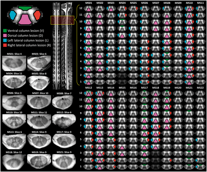
The study showed that the 7-tesla technique could pick up slight differences in functional connectivity in the spinal cords of 22 patients with a relapsing-remitting form of MS compared to healthy controls. Differences were most pronounced in regions of patients’ spinal cords that had visible lesions.
A lesion in the spinal cord presumably will cause some neurological damage. But this is the first time researchers have been able to noninvasively demonstrate that the presence of a lesion is correlated with altered neurological activity in the spinal cord.
These findings suggest that the technique might be used to determine whether medications, physical therapy or other interventions are preserving neurological function and thus slowing the course of the disease.
The research was supported in part by National Institutes of Health grants EB014841, NS092961, NS104149 and EB016689.