
A team of Vanderbilt University engineers and clinicians have won a five-year $1.8 million National Eye Institute grant to develop and translate novel intraoperative imaging technologies to the ophthalmic surgical suite to enable real-time surgical guidance.
“Our group has spent quite a few years working on developing the underlying imaging technology,” said Yuankai Kenny Tao, assistant professor of biomedical engineering and a faculty researcher in the Vanderbilt Institute for Surgery and Engineering, is the principal investigator. “This grant will support our translational efforts and allow us to identify ophthalmic surgeries that will directly benefit from real-time guidance and to better understand how surgical maneuvers impact postoperative visual function.”
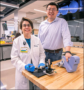
Photo: Anne Rayner|VU
Clinical collaborators at the Vanderbilt Eye Institute include Karen Joos, Joseph and Barbara Ellis Professor of Ophthalmology and Visual Sciences, professor of biomedical engineering and VISE researcher; Shriji Patel, assistant professor of ophthalmology and visual sciences; and Christine Shieh, assistant professor of ophthalmology and visual sciences.
Optical coherence tomography, a non-invasive imaging technique that provides high-resolution, cross-sectional images, is the “gold standard” for ophthalmic diagnostics. Cataract, glaucoma, age-related macular degeneration, corneal dystrophy and diabetic retinopathy are the leading causes of low vision and blindness in the United States and affect over 40 million Americans.
However, OCT-integrated surgical microscopy technologies have lagged and only recently have been FDA-approved and commercially available. Although recent studies have demonstrated the utility of iOCT for verifying completion of surgical goals, real-time feedback for ophthalmic surgical guidance is not feasible due to several fundamental limitations.
To address iOCT limitations, Tao’s multidisciplinary team of engineers and clinicians has developed 4D iSECTR (intraoperative spectrally encoded coherence tomography and reflectometry) technologies that offer real-time image data.
Surgery at advanced stages of eye diseases involves precision manipulation of delicate semi-transparent structures in the eye. Visualization of these tissue layers is critical to improving clinical outcomes and developing novel surgical techniques.
“We believe the imaging data from iSECTR will benefit surgical decision-making and lead to improved outcomes and the integration of imaging, registration, segmentation and feedback using heads-up display visualization will address unmet needs in conventional ophthalmic microsurgery,” Tao said.
“Collaboration among engineers and clinicians more efficiently enables the development of practical clinically useful devices and techniques,” Joos said.
The grant— Intraoperative Optical Coherence Tomography for Ophthalmic Surgical Guidance (R01-EY030490)—is administered through the National Eye Institute of the National Institutes of Health. Preliminary work leading to the award was supported in part by a VISE pilot grant, VISE physician-in-training grant, and the VISE NIH/NIBIB T32EB021937 training grant.