
With hundreds of thousands of people in the United States having a stroke annually, Vanderbilt researchers are developing technology that could revolutionize the way blood clots are removed by allowing surgeons to complete the process more efficiently and safely.
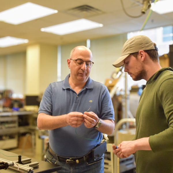
The research is led by Nabil Simaan, professor of mechanical engineering, with assistance from graduate student Jared Lawson. According to the Centers for Disease Control and Prevention (CDC), more than 795,000 people in the United States have a stroke every year. About 87% are ischemic strokes, in which blood flow to the brain is blocked.
The main objective of the surgeon, or interventionalist physician, is to find the clot causing the blockage and remove it. Currently, a thin flexible catheter is put into an artery in the patient’s groin and the surgeon uses X-ray images on a computer screen to move the catheter to where the clot is believed to be. Navigation is particularly challenging in the brain, researchers say, because of curving paths and tortuous vessels the catheter has to go through.
“An X-ray image doesn’t tell you where a blood clot is, it tells you where blood is flowing,” says Lawson. “So, they have to kind of decide, do I think the blood clot is here, or do I think it’s over here? That might add complications to the procedure, or it might just slow the procedure down.”
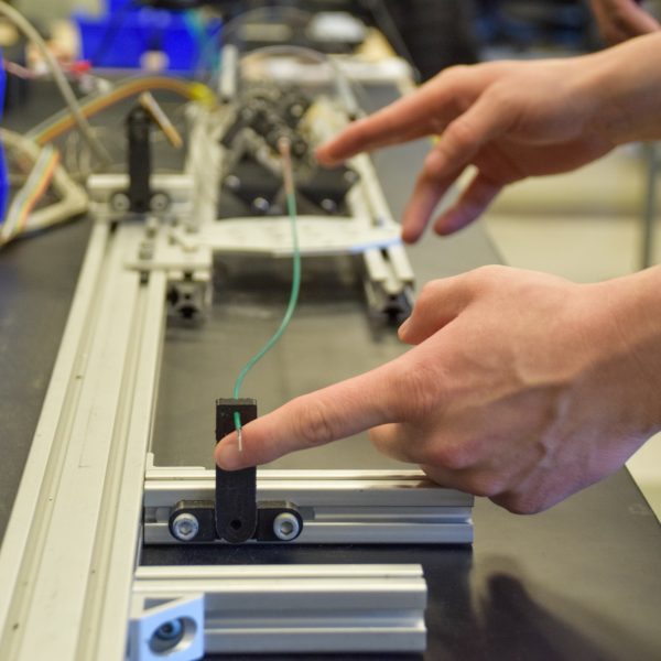
As a solution, Simaan and Lawson are working on adding sensors to aid navigation by allowing the surgeon to detect when the catheter tip is touching a vessel wall, and when it makes contact with a blood clot. The catheter, which is hollow, will also be equipped with a device that will pass through it and grab the clot or suction it like a vacuum.
“By informing the interventionalist of different contact scenarios, we hope to increase the safety and efficiency of these emergent neurovascular procedures,” says Lawson.
Additionally, he says they’re developing technology to automate steering by tracking the catheters motion under biplane X-ray fluoroscopy, which surgeons use to visualize the vessel anatomy.
“If we successfully automate the steering, the cognitive burden placed on the interventionalist can be significantly reduced,” he says.
Rohan Chitale, MD, is a clinical collaborator and associate professor of neurological surgery at Vanderbilt University Medical Center. He says surgeons currently use flimsy, passive catheters that require much trial and error.
“This new technology is revolutionary because it adds steerability, safety, and augmentation of senses that interventionalists do not currently have,” says Chitale, who is affiliated with Vanderbilt Institute for Surgery and Engineering (VISE), along with Simaan and Lawson. “Improving on these will make an interventionalist’s job simpler, safer, and more effective. Being able to have more certainty about where the clot is will mean more frequent and quicker success at restoring blood flow in the brain, thereby improving the outcome for countless stroke patients.”
The researchers say they appreciate the collaboration with Vanderbilt University Medical Center and point to it as another example of the partnerships the university fosters to produce top-notch research.
“To be able to work with a surgeon like Dr. Chitale, who actually performs these procedures, and get his feedback has been invaluable,” says Simaan. “This technology is groundbreaking in the sense that it can be used in any type of endovascular procedure, which means anything in the heart that would be catheter-based.”
Contact: Lucas Johnson, lucas.l.johnson@vanderbilt.edu