Medical images are a fundamental element in medical diagnosis and treatment, as they reveal the internal anatomy of patients. They contain information that human experts may not be capable of interpreting directly, and often medical image databases are too large to be effectively analyzed by humans. Medical image processing research involves developing algorithms and software that permit automatically or semi-automatically extracting critical information from medical image datasets. At Vanderbilt, research in the medical image processing field primarily focuses on development of machine learning and model-based image analysis methods for image feature extraction, segmentation, registration, and image-based bio-models. These methods have enabled numerous significant advances across the fields of medical science, including: (1) advancing medical knowledge through large scale analyses of images that inform structure and function of biological systems; (2) improving and creating new computer-assistance workflows for surgical procedures and other interventions that use information extracted from a patient’s medical images to customize the procedure; and (3) creating image-based computational models of biological systems to improve medical education and personalized treatment.
Benoit Dawant
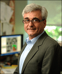 Cornelius Vanderbilt Professor of Engineering
Professor of Electrical Engineering, Computer Science, Biomedical Engineering, Radiology and Radiological Sciences, Neurological Surgery, Otolaryngology-Head and Neck Surgery
Director of the Vanderbilt Institute for Surgery and Engineering (VISE)
Benoit Dawant works at the interface between engineering and medicine with particular expertise in image processing and surgical guidance. Current projects focus on the development and clinical evaluation of algorithms and systems to assist in the placement of deep brain stimulators used for the treatment of Parkinson's disease and other movement disorders, the placement and programming of cochlear implants used to treat hearing disorders, the removal of brain tumors and the creation of radiation therapy plans.
For more than 10 years and with National Institute of Health funding, Dawant and Vanderbilt researchers have developed a system that facilitates the pre-operative, intraoperative and post-operative phases of deep brain stimulation (DBS) procedures. The system, which consists of a data repository called CranialVault and a suite of software tools called Cranial Vault Explorer, is the first computer-assistance system that spans the entire spectrum of the procedure.
In addition to his research, Dawant is at the helm of the Vanderbilt Institute for Surgery and Engineering, an interdisciplinary, trans-institutional institute whose mission is the creation, development, implementation, clinical evaluation, and commercialization of methods, devices, algorithms, and systems designed to facilitate interventional processes and their outcome. The center facilitates the exchange of ideas between physicians, engineers, and computer scientists. Primarily funded by federal research support, the center is also engaged with industrial partners for the commercialization of its intellectual property, the early evaluation of industrial devices and techniques, and the joint development of innovative solutions.
Catie Chang
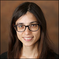
Sally and Dave Hopkins Faculty Fellow, Assistant Professor of Electrical Engineering, Computer Engineering,
Computer Science, and Biomedical Engineering
Catie Chang is principal investigator of the Neuroimaging and Brain Dynamics Lab (neurdylab) at Vanderbilt University. The goal of her research is to increase understanding of the human brain by advancing functional neuroimaging methods. Her lab develops novel approaches for uncovering information about brain activity in health and disease by combining functional neuroimaging (fMRI, EEG), signal processing, and machine learning techniques. Current project areas include: analyzing and visualizing the spatiotemporal structure of brain activity; multimodal imaging of dynamic brain states; and characterizing the neural and physiological underpinnings of fMRI signals. The research in her lab is interdisciplinary and collaborative, spanning fields such as engineering, computer science, neuroscience, psychology , and medicine.
Yuankai Huo
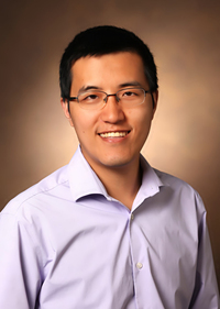 Assistant Professor of Computer Science, Computer Engineering
Yuankai Huo’s research interests (HRLB) include medical image computing, knowledge-infused machine learning, and large-scale healthcare data analytics. His research aims to facilitate data-driven healthcare and improve patient outcomes through innovations in medical image analysis as well as multi-modal data representation and learning. His current efforts focus on quantifying high-dimensional and spatial-temporal data from microscopy imaging techniques, including renal pathology, cancer pathology, cytology, computational biology. The quantitative imaging information is associated with molecular, genetic, and clinical features for precise diagnosis and treatment.
Bennett Landman
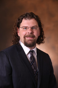 Chair, Department of Electrical and Computer Engineering
Professor of Electrical and Computer Engineering, Computer Science, Biomedical Engineering,
Radiology and Radiological Sciences, Psychiatry and Behavioral Sciences, Biomedical Informatics, Neurology
Bennett Landman leads the Medical‐image Analysis and Statistical Interpretation (MASI) lab at Vanderbilt University. His overarching goal is to combine image-processing technologies and electronic health data to improve understanding of individual anatomy and personalized medicine. His scientific tools focus on medical image processing with robust and scalable methods for large-scale data analysis. His primary training is in neuroimaging with a focus on diffusion weighted magnetic resonance imaging, and he has worked on Alzheimer’s disease and aging research since 2008. While his academic home is in electrical and computer engineering, Dr. Landman’s training is in biomedical engineering and 3-D radiological image analysis. He seeks to connect the image processing methods with both the medical physics underpinning of the data and their clinical applications. Dissemination and support of image processing tools is a core component of his lab’s interests, and his team constructed a university wide medical image processing system that handles data for 400+ IRB approved projects with more than 100,000 imaging sessions. Their system links closely with the university’s high-performance computing center for automated processing of structural, functional, and diffusion MRI data.
As an educator, Dr. Landman serves as Chair of the Department of Electrical and Computer Engineering and has directed the Electrical Engineering Graduate Program and served on the Graduate Faculty Council. He served as a Faculty Teaching Fellow, developed new laboratory course materials, and created new graduate courses in image processing. Dr. Landman is committed to committed to promoting rigorous and transparent research and supporting an inclusive academic environment.
Jack Noble
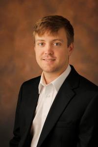 Assistant Professor of Electrical & Computer Engineering, Computer Science, Biomedical Engineering,
Head and Neck Surgery
Assistant Research Professor of Hearing and Speech Sciences (DHSS)
Director of Graduate Recruiting for Electrical and Computer Engineering
Jack Noble is the principal investigator for the Biomedical Image Analysis for Image-Guided Interventions Laboratory (BAGL). His lab investigates novel medical image processing and analysis techniques with emphasis on creating image analysis-based solutions to clinical problems. His research interests include state-of-the-art image analysis techniques, such as machine learning, statistical shape models, graph search methods, level set techniques, image registration techniques, and image-based bio-models.
With funding from the NIH, he is currently developing novel image analysis systems for cochlear implant procedures, including systems that use image analysis techniques for: (1) comprehensive pre-operative surgery planning through automated analysis of pre-operative CT scans, (2) intra-operative surgical feedback and guidance using intra-operative cone beam CT, and (3) development of patient-specific post-operative imaging-based electro-anatomical models of cochlear implant neural stimulation to assist audiologists with cochlear implant programming and improve hearing outcomes.