
Researchers from the Vanderbilt University Institute of Imaging Science (VUIIS) have detected signals in the white matter of the brain that suggest it has more neural activity than previously thought.
The findings of Zhaohua Ding, research associate professor of electrical engineering, computer science and biomedical engineering, and colleagues suggest a new way to investigate diseases such as Alzheimer’s and multiple sclerosis associated with a failure of white matter functional integrity.
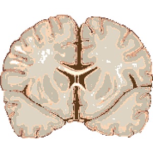
Until recently, bundles of long nerve fibers, or axons, that make up white matter were considered passive transmitters of signals between different brain regions. The gray matter contains neurons, or nerve cell bodies.
The researchers found synchronous BOLD signals in white matter reflecting neural activity both in a resting state and in response to functional loading. The BOLD (blood oxygenation level dependent) signal detected by functional magnetic resonance imaging reflects changes in the magnetic properties of blood as it transports oxygen to brain tissue.
Their results, published in the Proceedings of the National Academy of Sciences, support the notion that synchronous BOLD correlations representing functional connectivity are present in white matter and that neural activities are encoded in white as well as gray matter.
“While detection and characterization of BOLD signals, as well as their electrophysiological and hemodynamic/metabolic origins, have been extensively studied in gray matter, the detection and interpretation of BOLD signals in white matter remain controversial,” the authors wrote.
“In this study, we provide further strong evidence that BOLD signals in white matter reflect neural activities both in a resting state and under functional loading,” they said. “We demonstrate that BOLD signal wave forms in stimulus-relevant white matter pathways are synchronous with the applied stimuli but with various degrees of time delay and that signals in white matter pathways exhibit clear task specificity.”
The seven-member team included researchers as well as faculty members from VUIIS, the School of Engineering, the Vanderbilt Brain Institute, the Vanderbilt Kennedy Center, the Department of Radiology and Radiological Sciences, and Peabody College.
This research was supported by grants from the National Institutes of Health (NS093669, HD044073, HD067254, HD083211, HD090923).
Media Inquiries:
Bill Snyder, (615) 322-4747
william.snyder@Vanderbilt.Edu