
BIOMEDICAL IMAGING AND BIOPHOTONICS
Vanderbilt engineering researchers have developed a first-of-its kind ultrathin filter that processes images at the speed of light and supports direct imaging of an object’s boundaries.
Their work marks a significant breakthrough in using optics for image processing and holds transformative potential for applications in biological imaging and computer vision.
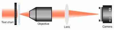
Digital image processing, including the use of neural networks, has become essential for identifying features and objects in images for many science and engineering disciplines, but it requires advanced computers, space to accommodate them, and substantial power to run them. Optical image processing actually predates its digital counterpart but requires multiple optical lenses and filters resulting in a large multi-optic system. The advance by the Valentine lab allows placement of a thin filter in front of a conventional camera for achieving the same functionality—significantly reducing the system’s size and complexity.
The Vanderbilt project demonstrated “two-dimensional image differentiators with high resolution, thin form factor and a simple geometry that allows rapid and cost-effective large-scale manufacturing,” said the team, led by Jason Valentine, associate professor of mechanical engineering. The work, “Flat optics for image differentiation,” was published online in Nature Photonics earlier this year.
“Optical analog processing has the advantages of being low power and high speed,” said Valentine, also deputy director of the Vanderbilt Institute of Nanoscale Science and Engineering.
The team’s filter is based on a two-dimensional photonic crystal made from silicon. It can be integrated into an optical microscope or onto a camera sensor, easily adapting an existing image processing system. The filter—100 times thinner than a human hair—also was integrated with a metamaterial-based lens, resulting in a completely flat, compact and ultrathin optic that can perform edge imaging.
“One of the primary benefits of our approach is the ability to integrate the metamaterial with traditional optical systems. As an example, we built an edge detection microscope by simply placing the metamaterial filter within a commercial optical microscope,” said You Zhou, a Ph.D. student in the Interdisciplinary Materials Science Program and one of the four authors.
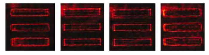
Testing included imaging the cells of onion epidermis, pumpkin stem and pig motor nerve and differentiating their boundaries, a task for which accuracy carries critical implications.
Edge filtering is a common preprocessing step in object recognition. “It is important, for instance, in detecting the edge of a lane for autonomous vehicles. It can also be used for detecting margins of tumors in medical imaging or in classifying cell size and type in the case of cell sorting for cancer detection,” Valentine said.
“The key feature is the ability to perform image processing at the speed of light while requiring no input power and doing so in an extremely thin form factor,” Valentine said. “This opens new doors for real-time and high speed optical analog image processing in applications such as machine vision and biological imaging.”
His group is now working to adapt the technology for use in object recognition, not just edge imaging. Those projects include more complex object recognition, funded by DARPA, and related technology for cell identification, which received internal Vanderbilt funding for projects with transformative potential.