
A significant component of the cost of an MRI system has been the massive superconducting magnet to produce a strong radiofrequency current and the bulky system that keeps it cool. The magnet for a 3-Tesla scanner, for example, weighs more than 12,000 pounds.
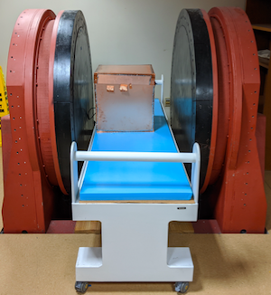
Vanderbilt engineers believe it’s time to downsize. A team led by William Grissom, associate professor of biomedical engineering, will use a very low-field human 47.5 millitesla scanner that it built, which is installed and working at the Vanderbilt University Institute for Imaging Science. With it as a testbed and a $1.4 million NIH grant, researchers will work toward a compact, silent, less expensive and potentially portable MRI device.
“That is 100 times weaker than normal research magnets,” he said. “Rather than weighing tons we are looking at a few hundred pounds.”
Researchers will develop new hardware, including low-field radio frequency transmission coils and amplifiers, and software that will together translate signals measured from the body into images of anatomy. And they’ll use new spatial encoding approaches that are completely different from those found on conventional clinical MRI scanners.
“The time is right to do this kind of work,” Grissom said. “Computational and electronic technologies have advanced so much over the last 40 years and become cheap enough that we can now look at whether we can get clinically useful images from less expensive and more portable magnets.”
The project builds on earlier work by the Grissom lab in coil design, spatial encoding using radiofrequency field gradients, and radiofrequency pulses. A key advancement will replace the type of gradient fields the scanner uses by taking advantage of the Bloch-Siegert shift, a phenomenon historically viewed as a nuisance in magnetic resonance. In this project, it will be used to match up signals received from the body to their location of origin and form images.
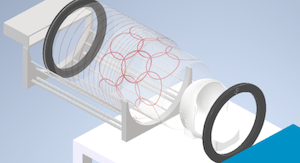
The gradient fields used now are problematic: they are loud and induce peripheral nerve stimulation, compromising patient comfort. They require bulky cooling systems and customized amplifiers. Together, that represents up to 30 percent of the cost of a clinical scanner.
Caption: Researchers will use a low-field MRI lab at VUIIS in developing new hardware and software for a less expensive and more portable system. They also will use new spatial encoding approaches.