
The relatively brief history of medical MRI is riddled with failed predictions, according to University Professor John Gore, founding director of the Vanderbilt University Institute of Imaging Science.
Bold statements about the optimal magnetic field and the limits of magnet strength were way off. In 1982 one researcher concluded MRI was useful for imaging the human body but not the brain because CT scans did it better. That same year another researcher said MRI’s biggest advantage over CT was that it did not need contrast agents. Wrong and wrong.
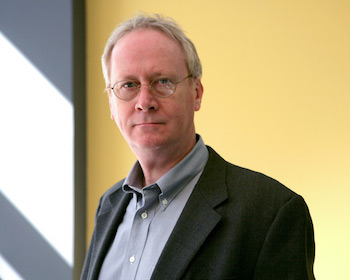
An MRI pioneer himself, Gore talked about the imaging method’s history, state of art, and future directions last month at the 2021 annual meeting of the International Society for Magnetic Resonance in Medicine. He was selected to deliver the Paul C. Lauterbur Lecture, the most prestigious lecture in magnetic resonance imaging. Lauterbur was a U.S. chemist who shared the 2003 Nobel Prize in Physiology or Medicine with Peter Mansfield for work that made development of MRI possible.
The Lauterbur Lecture is the main scientific talk that concludes the annual ISMRM meeting each year, and this year, a virtual conference, it drew 5,500 participants.
Will Grissom, associate professor of biomedical engineering, is on the ISMRM Annual Meeting Program Committee and introduced Gore for the lecture. His introduction highlighted Gore’s role in the birth of MRI, his contributions to contrast-enhanced, microstructural, and functional MRI, and his establishment of the Vanderbilt University Institute of Imaging Science.
“It was a great honor to be asked to give the Lauterbur lecture and an opportunity for me to recall some of the highlights of the early days of MRI and my encounters with Paul Lauterbur himself.”
The scientific principle behind MRI, magnetic resonance imaging, was developed in the 1940s but used mainly to study the chemical structure of substances until the 1970s. With advancements by Lauterbur and Mansfield, it could be used to produce images of the body, organs and tissues.
“Technical innovations have dramatically improved image quality–including resolution, speed, and signal-to-noise ratio–but unlike CT, our understanding of how to explain MRI images precisely based on tissue properties lags behind,” said Gore, professor of radiology and radiological sciences, biomedical engineering, molecular physiology and biophysics, and physics and astronomy.
“We need more complete, validated models of MRI contrast to allow the full impact of efficient acquisition and quantitative parameters, and this may lead to more novel types of information from MR images,” said Gore, who also is the Hertha Ramsey Cress Chair in Medicine.