
By Leah Mann

Researchers in the labs of Laurie Cutting, Patricia and Rodes Hart Professor and professor of special education at the Peabody College of education and human development, and Bennett Landman, professor and chair of the Department of Electrical and Computer Engineering at the School of Engineering, recently published a study in Magnetic Resonance Imaging exploring a new tool for imaging the brains of neurofibromatosis type 1 patients and its significance in assessing their symptoms.

Neurofibromatosis 1 is a disorder caused by mutations in a gene that regulates the production of a tumor suppressor. These mutations can trigger the growth of tumors such as neurofibromas, benign tumors that develop on nerve cells.
The best way to see many of the features of the disease is to do FLAIR imaging (a type of magnetic resonance imaging) on the brains of patients. In FLAIR images of neurofibromatosis 1 patients, there often are abnormally bright spots that likely indicate the presence of fluid between layers of myelin sheaths—the insulation surrounding certain nerve cells—which may contribute to the cognitive deficits observed in patients.
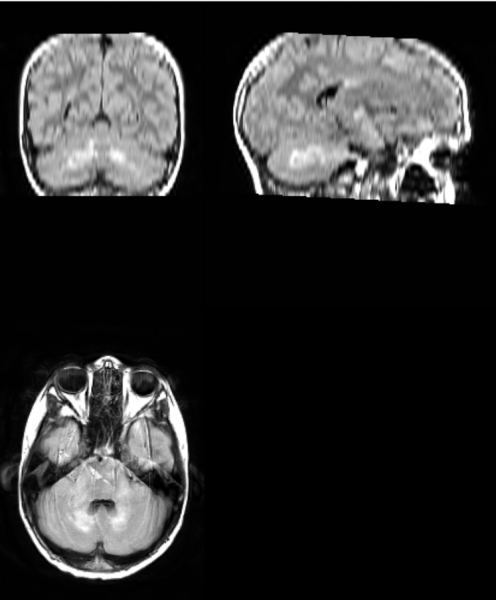
Unidentified Bright Objects—the bright spots in the FLAIR images—have been traditionally defined and segmented (digitally separated for further study) manually, which requires intensive training and is time-consuming and expensive. The current work, led by first author and neuroscience Ph.D. student Emily Harriott and co-author and recent neuroscience Ph.D. graduate Tin Nguyen, tested and validated a semi-automated approach for detecting and segmenting UBOs in children and adolescents with neurofibromatosis 1. Cutting lab staff researcher Laura Barquero also participated in the study.
The new approach, which uses a tool called the Lesion Segmentation Tool, reliably identified and segmented UBOs and was much faster than doing the work manually. After establishing the utility of LST, the scientists explored possible correlations between total UBO volume and cognitive function. The researchers demonstrated that total UBO volume was related to single word reading ability, phonological awareness (recognizing and manipulating spoken parts of speech) and visuospatial skills, and participants with greater UBO volumes had lower scores on these cognitive measures.
Using automated tools will decrease the time and cost needed to analyze UBOs. Additionally, applying tools like LST to examine the relationships between UBO volumes and cognitive abilities will help create and refine targeted pharmacological, academic and behavioral interventions that may improve neurofibromatosis 1 patients’ quality of life.
Go Deeper
The article “Using a semi-automated approach to quantify Unidentified Bright Objects in Neurofibromatosis type 1 and linkages to cognitive and academic outcomes” was published in Magnetic Resonance Imaging in January 2023.
Funding
This study was funded by the National Institutes of Health, the National Center for Advanced Translational Science, the Eunice Kennedy Shriver National Institute of Child Health and Human Development, the National Institute of Neurological Disorders and Stroke, and the Mind Science Foundation.