
New applications of artificial intelligence (AI) to renal pathology have been driven by the widespread use of digital diagnostic imaging and interdisciplinary collaborations between computer scientists, nephrologists and renal pathologists with potential for major impacts in diagnosis and understanding of kidney diseases.

A Vanderbilt computer scientist is working with key clinical collaborators at Vanderbilt University Medical Center to develop a quantitative and reproducible 3D analytics tool for large-scale digital analysis of kidney tissues and biopsies.
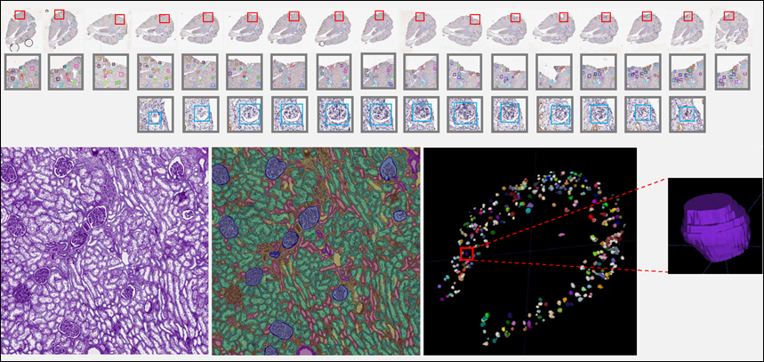
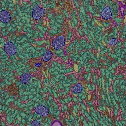
“Our research aims to equip clinical scientists with an advanced 3D spatial and 3D cellular-level analytics tool to investigate the anatomical-molecular association in kidney tissues,” said Yuankai Huo, assistant professor of computer science and electrical and computer engineering, and faculty affiliate in the Vanderbilt Institute for Surgery and Engineering. The short-term goal is to enable the AI-empowered 3D computer vision on routine digitized renal tissue biopsies that will allow renal pathologists to perform a reproducible 3D phenotyping on 2D whole slide images, which are small high-resolution image strips combined to create a full image of a tissue section.
For this project—AI-empowered 3D Computer Vision and Image-Omics Integration for Digital Kidney Histopathology—Huo has received a five-year, $2.7 million grant from the National Institutes of Health. The physician collaborators are Haichun Yang, Agnes B. Fogo and Shilin Zhao. Yang is a research assistant professor and Fogo is a professor in the Department of Pathology, Microbiology and Immunology at Vanderbilt University Medical Center. Zhao is an assistant professor in the Department of Biostatistics at the medical school.
“The success of the project will pave the way for a fully quantitative, high-throughput and reproducible 3D image-molecular analytics (spatial-transcriptomics) for medical image computing and bioinformatics communities, two essential research fields in computer science,” Huo said.
“I am grateful for a Vanderbilt Institute for Surgery and Engineering seed grant that has helped to fund the pilot data collection phase of our project. As an early-career PI, VISE’s support has played a crucial role in building strong relationships between myself and our collaborators.” Huo said.
The research is supported by a National Institutes of Health R01 grant DK135597-01