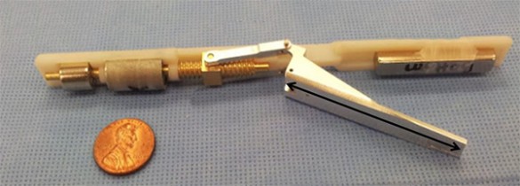
Pietro Valdastri is convinced that the clever application of magnetic force can make minimally invasive surgery easier and more effective.
“In 2007, a team of University of Texas researchers did some basic experiments using magnets in laparoscopic surgery,” said Valdastri, assistant professor of mechanical engineering and director of Vanderbilt University’s Science and Technology of Robotics in Medicine (STORM) Lab.
“Although their designs were very simple, mechanically speaking, they made me realize that small surgical devices guided and powered by external magnets have a number of potential advantages over placing tools on the end of a stick, which is the current approach. All that was required is a little sophisticated engineering!”
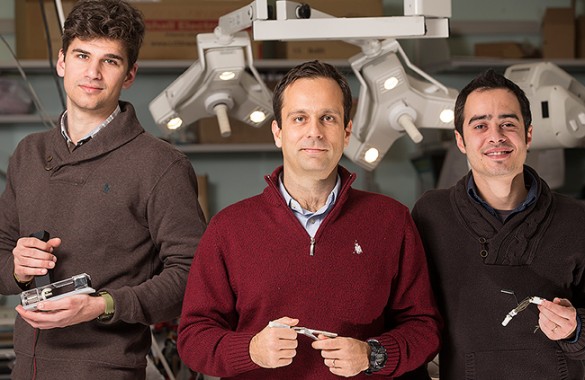
The researchers who are developing magnetically driven laparoscopic instruments: Nicolò Garbin, left, Piero Valdastri, and Christian Di Natali. (Joe Howell / Vanderbilt)
This realization led Valdastri and his graduate students – particularly Christian Di Natali and Nicolò Garbin – to develop a new approach to laparoscopic surgery that they call local magnetic actuation (LMA) and describe in an article titled “Closed-Loop Control of Local Magnetic Actuation for Robotic Surgical Instruments” published recently in the journal IEEE Transactions on Robotics.
The approach requires two components: an external unit that is placed on a patient’s abdomen and an internal unit small enough to fit through the ¼ to ½ inch ports that are used in minimally invasive surgery. Each component carries a set of two strong permanent magnets. One pair anchors the internal unit to the inside of the abdominal wall, while the other pair provides the mechanical force that powers the device.
According to the researchers, their approach has two major benefits over the current “tool on a stick” approach. The most important is that it can deliver between 10 to 100 times more mechanical power than the electrical motors that are small enough to fit through the surgical ports can provide. The second is that it is easier to place the magnetic devices at optimal positions within the body than it is to position devices connected to sticks or wires.
The first instrument that embodies this principle is a fountain-pen-sized magnetic organ retractor that is described in an article appearing in the March issue of the ASME Journal of Medical Devices.
Di Natali came up with the innovative design, which Garbin then developed as a visiting master’s student from the Polytechnic of Milan. After getting his master’s degree, he joined STORM Lab as a doctoral student and continued working on the device.
The device is designed to move organs out of the way when needed to perform an operation. For instance, the liver lies on top of the gall bladder, so it must be moved aside before the gall bladder can be removed. That isn’t much of a problem in old-fashioned open surgery, but open surgery is increasingly being replaced by minimally invasive surgery where the operation is performed with special equipment through small incisions in the abdominal wall.
Although patients tend to have quicker recovery times and less discomfort with minimally invasive surgery than with conventional surgery, being forced to operate through small incisions (a procedure aptly nicknamed keyhole surgery) raises a new set of issues for the surgeon. One of these is organ retraction.
As it is currently practiced, minimally invasive abdominal surgery frequently requires making three to five incisions in order to insert all of the equipment needed. When an organ must be repositioned, an extra incision is needed to insert the organ retractor. One type of retractor looks like a miniature garden rake. The tines are collapsed together so they will fit through the incision and then spread apart so they can grip an organ with enough force to shift its position. The range of movement of the retractor is limited by the location of the incision through which it is inserted.
The STORM device looks much different than a conventional retractor. It consists of two parts: a fountain-pen-sized unit that is inserted into the body and a larger external unit that is placed on the patient’s stomach. (There is no need to make an additional incision because the device can be inserted through a port used by another instrument.)
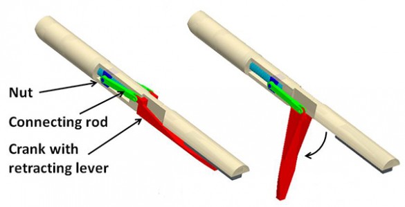
A computer drawing of internal retractor unit, which is about the size of a small fountain pen. (STORM Lab / Vanderbilt)
Both units contain powerful permanent magnets that are oriented so they attract one another. The magnetic force is strong enough to firmly anchor the internal unit against the inside of the abdominal wall directly below the external unit. The magnetic attraction between the two allows the user to accurately position the inside unit by rotating and sliding the external unit.
Both units also contain a second set of magnets that transmit the power. The magnet in the external unit is attached to the shaft of a powerful electric motor that causes it to spin. The magnet in the internal unit is also attached to a shaft, but one that drives a two-inch lever. When the electric motor on the external unit twirls its magnet, it generates a rotating magnetic field that forces the magnet on the shaft in the inner unit to spin at the same speed. When it spins in one direction, the lever opens up and when it spins in the opposite direction, the lever closes.
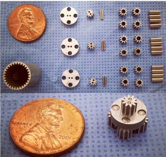
To make the retractor small enough to fit through the incisions used in minimally invasive surgery, the engineers had to create a drive train with a number of very small gears. (STORM Lab / Vanderbilt)
To retract an organ, the surgeon picks up the internal unit with a laparoscopic grasper and inserts it through the port into the body. When he maneuvers the internal unit close enough to the external unit, it snaps into position against the inner surface of the abdominal wall. The motor on the external unit is engaged, lowering the lever. Using standard laparoscopic instruments, the surgeon attaches one end of a line to the tip of the lever and the other end to a clip or suction cup fastened to the organ that must be moved. The electric motor is run in reverse and the lever retracts, pulling the organ into the desired position.
Valdastri and his colleagues have tested the device in the lab and found that it can lift a 500-gram (1.1 pound) weight without any problem. The internal unit remained anchored and the lever opened and closed smoothly. They also tried it out with pig livers, which are comparable in size to human livers. Although the livers typically weigh three to four pounds, the device was able to lift and hold up the portion that typically covers the stomach and gall bladder with power to spare. The device has successfully undergone large animal testing.
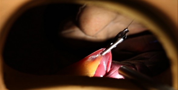
Demonstration of the power of magnetic actuation as magnetically driven retractor lifts the edge of a liver. (Joe Howell / Vanderbllt)
“This device demonstrates for the first time that controllable mechanical power can be transferred across the abdominal wall via an intelligent magnetic link to power a robotic instrument,” said Valdastri.
In the future, the engineer intends to apply this same principle to creating more complex instruments, such as laser and radio-frequency scalpels.
Besides the ability to deliver a lot of power, the magnetic actuation approach has some other important advantages: the internal units do not contain any expensive and delicate electronics so they can be easily sterilized and, if manufactured in bulk, could be made inexpensively enough to be disposable, Valdastri said.
One of the device’s main limitations, Valdastri acknowledged, is that it does not work if a patient’s abdominal wall is too thick. The magnetic force between the inner and outer units weakens rapidly as the distance between them increases and, at about an inch, it becomes too weak to keep the inner unit firmly anchored. As a result, the magnetic retractor will not work with overweight adults, which means about a third of the population in the U.S.
To turn this limitation into a strength, Valdastri’s group is adapting this approach for pediatric surgery and have begun developing a pediatric camera.
“Currently, no one has developed laparoscopic devices specifically for pediatric surgery,” Valdastri said. “There is a real need that I think we can help fill.”
Jacopo Buzzi and Elena De Momi from the Polytechnic of Milan and Vanderbilt doctoral student Marco Beccani also contributed to the research, which was funded by National Science Foundation grants 1239355 and 1453129.
Contact:
David Salisbury, (615) 322-NEWS
david.salisbury@vanderbilt.edu
